PCL Avulsion Injuries Anatomy is a critical topic in orthopedic medicine, as these injuries involve the detachment of the posterior cruciate ligament (PCL) from its bony attachment, typically at the tibia. The PCL is one of the key stabilizing ligaments of the knee, responsible for preventing the tibia from moving too far backward relative to the femur. Understanding its anatomy and the nature of avulsion injuries is essential for accurate diagnosis, effective treatment, and optimal recovery.
Anatomy of the Posterior Cruciate Ligament (PCL)
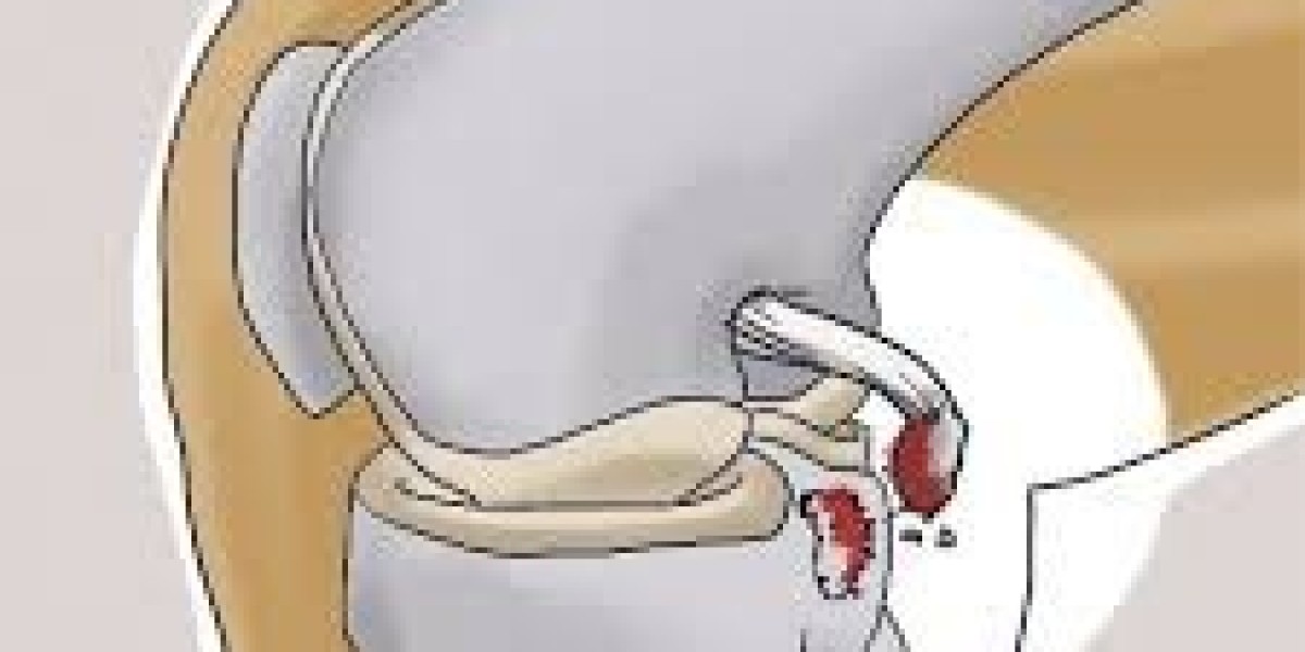
The PCL is located in the center of the knee joint, running from the posterior intercondylar area of the tibia to the medial condyle of the femur. Unlike the anterior cruciate ligament (ACL), the PCL is stronger and thicker, allowing it to withstand significant forces. Its primary functions include:
Posterior stability: Prevents backward displacement of the tibia.
Rotational control: Assists in stabilizing internal and external rotation of the knee.
Load distribution: Helps absorb stress during weight-bearing and dynamic movements.
The PCL is composed of two main bundles:
Anterolateral bundle – Tight in flexion, resists posterior tibial translation when the knee is bent.
Posteromedial bundle – Tight in extension, provides stability when the knee is straight.
What Are PCL Avulsion Injuries?
A PCL avulsion injury occurs when the ligament pulls away from its attachment on the tibia, often taking a fragment of bone with it. This type of injury is less common than mid-substance PCL tears but is particularly significant because it can destabilize the knee and impair function if untreated.
Causes of PCL Avulsion Injuries:
Trauma: High-impact injuries such as car accidents or sports collisions.
Hyperflexion or hyperextension: Sudden extreme knee movements can avulse the ligament.
Direct blow to the tibia: Often seen in dashboard injuries during car accidents.
Clinical Presentation
Patients with a PCL avulsion injury may experience:
Posterior knee pain and swelling
Difficulty walking or bearing weight
A feeling of instability, especially when descending stairs
Positive posterior drawer test on physical examination
Imaging studies, including X-rays, CT scans, and MRI, are crucial for confirming avulsion fractures and assessing the size and displacement of the bony fragment.
Treatment Approaches
Treatment depends on the severity and displacement of the avulsed fragment:
Non-surgical management:
Indicated for minimally displaced fragments
Includes bracing, physiotherapy, and gradual return to activity
Surgical intervention:
Recommended for displaced fragments or cases with significant knee instability
Techniques include open reduction and internal fixation (ORIF) or arthroscopic fixation
Early surgical repair often results in better functional outcomes
Rehabilitation and Recovery
Post-treatment rehabilitation focuses on restoring range of motion, strength, and stability:
Initial phase: Reduce swelling and protect the repair with a brace
Intermediate phase: Gentle range-of-motion exercises and quadriceps strengthening
Advanced phase: Functional training, proprioception exercises, and sport-specific drills
Recovery typically ranges from 3 to 6 months, depending on the extent of injury and treatment approach.
Conclusion
Understanding PCL Avulsion Injuries Anatomy is essential for healthcare professionals and patients alike. The PCL plays a vital role in knee stability, and avulsion injuries, though less common than ligament tears, require timely diagnosis and appropriate treatment to prevent long-term complications. Early intervention, whether surgical or conservative, combined with a structured rehabilitation program, can restore knee function and enable patients to return to their regular activities safely.